Written by Arthur Watson, NECO
Background/Intro:
Since starting my fourth-year rotations, there have been many learning experiences! While rotating through a Veterans Affairs (VA) eye clinic, I saw a unique patient whose care required multiple skills across my previous three years of learning. This case highlights the importance of individualized care, the need for adaptability and the necessity for the basic skills of the ophthalmic exam.
Case Presentation:
A 59-year-old Asian male presented to the optometry clinic with a chief complaint of blurry vision at near. He also reported a secondary complaint that his over-the-counter (OTC) reading glasses were sliding down his nose and frequently needed to be pushed back up. His change in vision was bilateral, gradual and was mostly relieved with the OTC readers when properly placed.
This was his first visit to the optometry department. His ocular history was significant for LASIK OU performed 16 years ago. The patient was unsure of prior refractive error but reported being significantly nearsighted. His past medical history was significant for traumatic quadriplegia with its associated sequelae but was otherwise noncontributory. He was accompanied by a caretaker to the exam room. He currently uses artificial tears as needed for dry eyes, which are instilled by his caretaker. The caretaker is also who adjusts his glasses when needed.
Family ocular and medical history were both unremarkable.
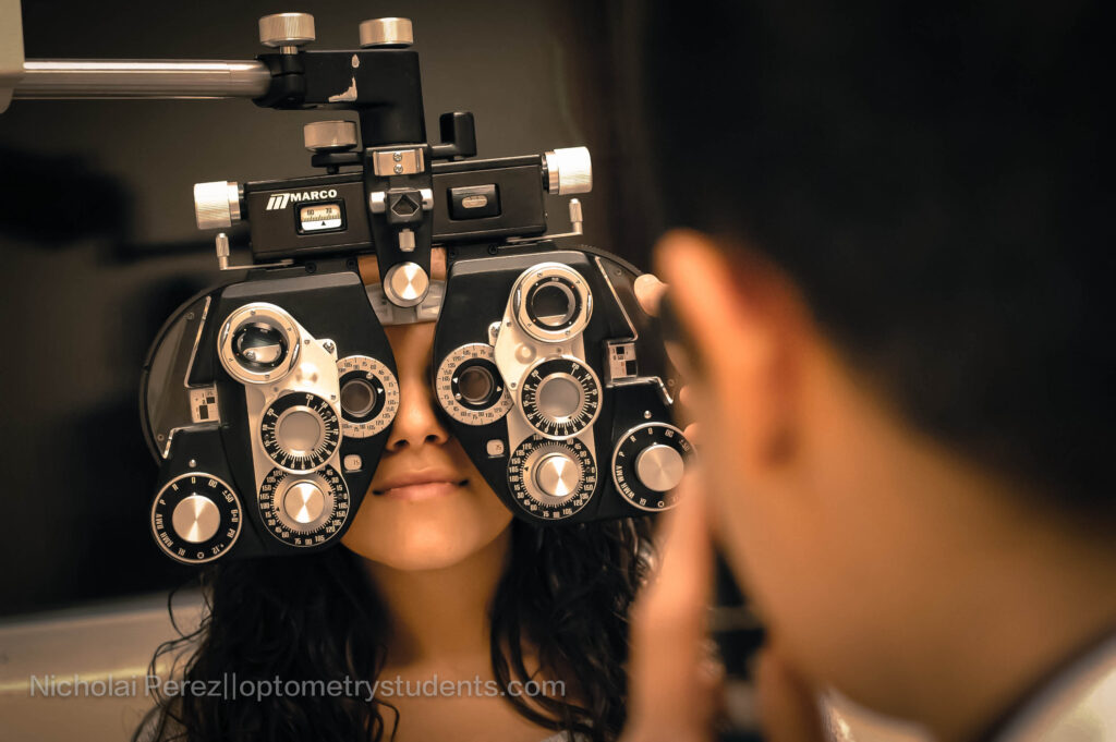
Upon examination, pupils were equal, round and reactive to light OU and without an afferent pupillary defect. Extraocular motilities were full and smooth OU. Confrontation fields were full to finger counting OD and OS. Retinoscopy was performed to obtain a starting point. Manifest trial frame refraction yielded a refractive error of OD: +2.00-1.00×010 and OS: +0.75 spherical with an add of +2.50 OU. Visual acuities were 20/20 OD and OS at distance and near.
Slit lamp examination was performed with a handheld slit lamp OD, and a 20D lens with a light source OS; this difference was because the handheld battery died. Examination of the anterior segment was remarkable for post-LASIK scars OU and mild complexion-associated melanosis OD. Intraocular pressure with TonoPen was unreadable OD and 6mmHg OS. Using digital pressures, the globes were soft and equal OU. The patient was dilated with one drop each of 2.5% phenylephrine and 1% tropicamide OU.
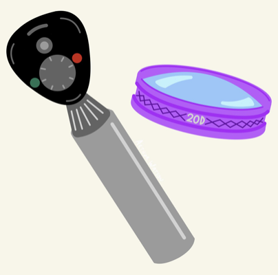
Examination of the posterior segment was done with a binocular indirect ophthalmoscope and a 20D lens and was remarkable for few mid-peripheral intraretinal hemorrhages OD and vitreal syneresis OU. Posterior pole evaluation was performed with BIO and a 15D lens as well as with a direct ophthalmoscope and was unremarkable for any optic nerve or macular irregularities OU.
Diagnosis & Plan:
The patient was diagnosed with anisometropic hyperopia OD>OS, regular astigmatism OD, and presbyopia. Given that the patient had only been wearing OTC readers, we decided that the best first line treatment would be spectacle correction with a multifocal lens. The patient declined this choice and preferred using single vision near correction. Due to the patient’s age and because these would be the first glasses since prior to his LASIK surgery, we decreased the manifest hyperopia OU to avoid any possible aniseikonia issues while keeping the manifest astigmatism OD. Final near vision only prescription was OD: +3.50-1.00×010 and OS: +3.00 spherical. We educated the patient that a properly made and fit pair of glasses would hopefully reduce the issue of his glasses sliding down. The patient reported that this was a significant burden to his quality of life and his lack of limb use necessitated him asking for assistance from his caretaker every time they slid down. We encouraged him and discussed other possible treatment options with contact lenses such as multifocal lenses or a monovision correction if his newly fit glasses were ineffective. He was satisfied with this treatment plan and a follow up was scheduled to evaluate the effectiveness after he had received his glasses and had them fit.
Additionally, upon further history taking, the patient reported he had recently had a considerable episode of sneezing, which he reported to be quite severe. Considering this, his lack of vasculopathic risk factors and negative blood thinner history, he was diagnosed with peripheral retinal hemorrhages OD likely due to valsalva. He was educated and a follow-up was scheduled to evaluate for resolution.
Conclusion:
This patient case displays a few challenges, which necessitated changes to what was the “typical” patient work up in a VA setting.
Examination of the external structures needed to be performed using a handheld slit lamp as well as a condensing lens and a light source. The handheld slit lamp is preferable for improved visualization and tissue evaluation, but due to the battery dying during the examination, the quick alternative of a light source and a 20D lens had to be chosen. Additionally, not all clinicians may have a handheld slit lamp at their disposal. Therefore, the knowledge of the less ideal alternative is still valuable.
Intraocular pressure measurement with TonoPen was only possible in one eye. Using that baseline and then digital pressures to compare equality was the only available method. Although non-ideal, this comparison and evaluation is more meaningful than recording the inability to perform TonoPen or Goldmann applanation. This would have been a good patient to use rebound tonometry such as the iCare, but this was not available.
Comfort with the trial frame refraction is valuable in scenarios such as this one, where a patient cannot reasonably be placed behind a phoropter. Additionally, this patient reminded me that retinoscopy is not only for pediatric providers! This skill is a quick and valuable means of obtaining a starting point for a patient when autorefraction is not possible and there is no prior data available.
Ophthalmic examination was mildly modified. We utilized a lower dioptric power condensing lens (15D) with the BIO, which affords a more magnified view of the posterior segment with the associated decrease in visual field. Additionally, we also evaluated the posterior segment with direct ophthalmoscopy. Both of these changes are simple but not commonly done with the routine patient who can easily be examined with a 20D lens or at the slit lamp with a 90D lens. It’s important to remember these options for thoroughly evaluating a patient such as the one described here. Additionally, comfortable use of the direct ophthalmoscope is a skill that should be easily performed by an eye care provider. I have found the increased magnification very beneficial when evaluating subtleties of the optic nerve. While on rotations, I have used the ophthalmoscope to help delineate disc hemorrhages that are very subtle on both 90D exam as well as fundus photographs.
In this case, we addressed the patient’s complaints of near vision blur and unstable OTC readers by prescribing single vision only spectacles and emphasizing the importance of properly fit frames. It is important to consider alternative methods for clear near acuity especially in unique cases like this patient without limb function. Continually adjusting their glasses may be a detriment to their quality of life or may be entirely impossible. Options discussed with this patient were differing contact lens modalities. Other potential options for a patient like this include Vuity ophthalmic drops or surgical treatment. Possible surgical options could be a clear lens exchange with a multifocal intraocular lens or possibly a Kamra corneal inlay. All of these options have their disadvantages and potential side effects. A risk versus benefit assessment must be done.
In conclusion, patients deserve comprehensive evaluations despite potential physical ailments.
It’s imperative to be flexible and inclusive while still upholding the highest standards of care and creating a safe, comfortable environment for the patient. It is necessary to know and be comfortable with the basic examination methods so that when scenarios like these arise, we can have reliable measurements and veridical evaluations. As future eye care providers, it is important to remember the tools and tricks within our clinical armamentarium! Finally, unique patients require unique treatment plans, and we should care for each patient individually. It’s valuable to consider the patient’s needs, their complaints, and formulate a treatment plan that is individualized and the patient is agreeable to.