Age/Sex/Race
2 year old Caucasian female
Chief Complaint
“My granddaughter’s pediatrician thinks her eyes aren’t working together and wants her checked for a lazy eye. It only happens sometimes in the left eye.”
Medical History / Ocular History / Family History
Unremarkable
Medications
None
NKDA
 Diagnosis and initial plan of action
I thought it might be an eye turn or a refractive error.
Applicable Testing & Results of Testing
Distance visual acuity (uncorrected): Unable to obtain OU
Near visual acuity (uncorrected) 20/80 with Lea chart OU
Cover test: ortho at distance and near
Extraocular muscles: grossly full
Pupils: PERRLA OU, (+) APD OD
Angle Kappa: +0.5mm nasal with white reflex OD, +0.0 mm red reflex OS
MEM:
OD: No reflex
OS: +0.50 -0.50 X 180
Slit lamp examination
Anterior segment: unremarkable
Lens: clear OD with white structure right behind it, clear OS
Dilated Fundus Examination:
OD: White structure with vasculature
Unable to see past the structure with BIO, Fundus lens or o-scope
OS: Glimpse of ONH with O-scope
 Assessment and Plan
We diagnosed our patient with retinoblastoma OD. We had them go see a pediatric ophthalmologist within a week. It was so large that we assumed that she would need an enucleation. A pediatric ophthalmologist confirmed that it was a retinoblastoma and she will need enucleation. However, we did not find out whether it metastasized anywhere else. There was no family history of retinoblastoma. If the pediatrician didn’t recommend her to come in, she would have never discovered it until it was too late.
Here’s some more information about retinoblastoma:
It is the most common primary, intraocular malignancy in children. It accounts for about 3% of all childhood cancers. Retinoblastoma occurs due to mutation in RB1, a tumor suppressor gene, in retinal cells. If one sibling is affected, there is a 2% risk that another sibling will be affected. If parents are affected, there is 40% risk that a child will inherit it. About 60% of cases are heritable and unilateral. A child will often present with leukocoria, strabismus, secondary glaucoma, poor vision, pain or redness around the eyes. In severe cases, orbital inflammation, proptosis and retinal detachment can occur. Ultrasound, CT or MRI can be used to aid in diagnosis of retinoblastoma. Depending on the size of the tumor, different treatment options can be used such as photocoagulation, cryotherapy, brachytherapy, external beam radiotherapy, chemotherapy or enucleation.
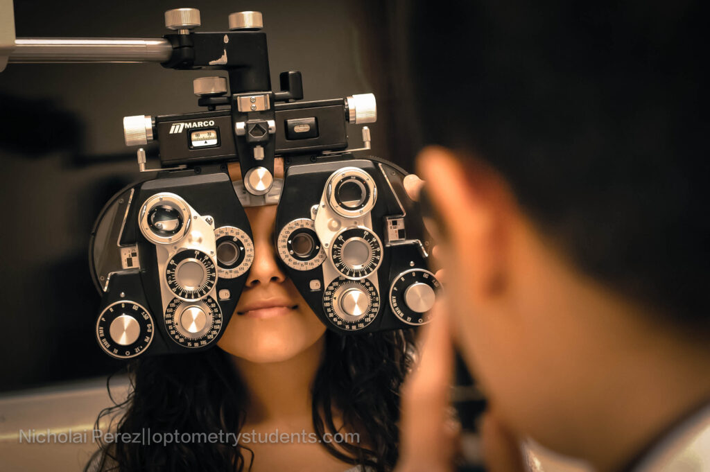
**Picture courtesy of Nicholai Perez of OS.***
References:
http://radiopaedia.org/articles/retinoblastoma
Tasman, W. & Jaeger, E.A. Atlas of Clinical Ophthalmology , 2nd edition. Lippincott, Williams & Wilkins, 2001.
Kanski, J. Clinical Ophthalmology A Systematic Approach. Elsevier, 6th edition, 2011.