One of the hardest things in optometry school can be getting comfortable with performing different skills and using the various equipment with ease during an eye exam. Something that has really taken off is the use of virtual reality in the education setting.
Virtual reality as a study tool
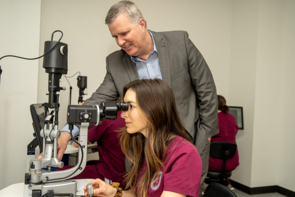
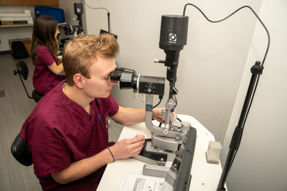
Here at The Kentucky College of Optometry (KYCO), we have direct 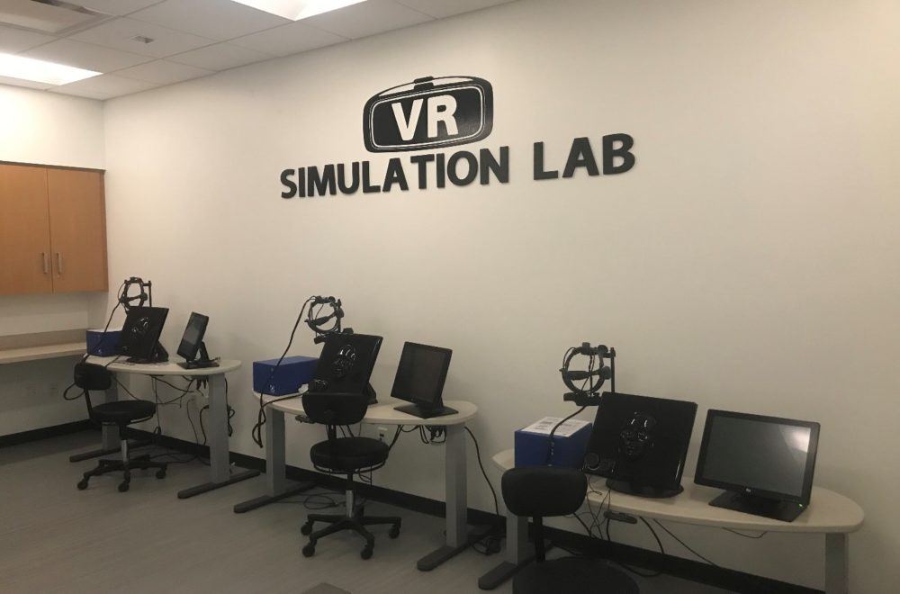 ophthalmoscope, bionocular indirect ophthalmoscope, and now the slit lamp VR Magic simulators. If you’re unfamiliar with VR Magic, this advanced technology is helping optometry students practice clinical skills, well…virtually! The specialized equipment looks and feels almost exactly like the equipment used when examining a patient in the exam room; the BIO headsets adjust just like a standard headset would, the ophthalmoscopes have the same power adjustment knobs that you would find on an actual direct ophthalmoscope, and the slit lamps have a realistic feel with how it moves for a better handle on lighting adjustments and handling. One great thing about these virtual reality systems is that they all connect to a computer monitor that allows you to keep track of certain areas you have looked at and you can even take screenshots of certain views or angles to reference later.
VR Slit Lamps
The Kentucky College of Optometry is the first in the world to receive the VR Magic Slit Lamp/Biomicroscope simulators. When looking through the oculars, you are looking into a virtual patient’s eye and perform a slit lamp exam just as you would on a patient in your exam chair. Most of these VR Magic programs have specific pathology that you have to view and record and it does this in a manner that gives you a grade. This is a game-changer when it comes to mastering skills because it can be done on your own time.
This means no more asking another student or volunteer to sit as you shine bright lights around to learn the ropes! Our clinical skills professors have noted that, overall, the students seem more confident and knowledgeable approaching a new skill when having practiced on a simulator before performing the skill in a methods lab.
Enhanced Learning
Through training and test programs on the software, you can learn how to grade different severities of a cataract or cells and flare. This is extremely useful to see a comparison of different stages of cataract formation since most optometry students don’t have any cataracts to observe in lab.
It can be intimidating knowing that you may only see a certain pathology on a picture or, even scarier, when you’re actually working in clinic. One of the 4th-year students said that she wished she could have practiced on the VR slit lamp before getting into clinic because even though class notes are helpful, it’s one thing to read a description of what you look for in certain pathology, but it’s another thing to see it “in action” right in front of you. This is why it’s so advantageous to have a training software like VR Magic to be exposed to, especially because there are simple pathological findings that you need to be able to assess starting day 1 of clinic, such as stages of diabetic retinopathy or macular degeneration.
Take advantage
Even with today’s technological advancements, using virtual reality programs such as VR Magic has to be at just about the top of the list when it comes to technology that will really help you get through optometry school. The slit lamp is an essential tool for a comprehensive eye exam, making it imperative that optometry students become masters of this skill as early into the program as possible.
If your school has invested in VR Magic technology or if you have the opportunity to use in it in the future, I believe students should take advantage of this amazing technology to help build not only the clinical knowledge of eye pathology, but to build confidence in operating the equipment, too.
