Background: The embryonic fissure of an embryonic eye is located slightly nasally and inferiorly, extending from the optic nerve to the pupillary margin. A coloboma is defined as a defect in one or several of the ocular structures due to the incomplete closure of the embryonic fissure. A coloboma can be unilateral or bilateral and usually occurs sporadically in healthy individuals; however, there are various associations with an optic disc coloboma. The systemic conditions associated with a coloboma include: chromosomal abnormalities, CHARGE syndrome, Goldenhar syndrome, and central nervous system abnormalities. The autosomal dominant variety is caused by the PAX6 gene mutation. (1)
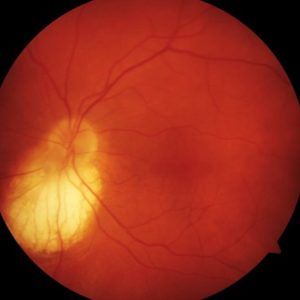
Signs: VA is often decreased, with the potential of amblyopia and refractive error. There will be a focal, glistening, white, excavation that is decentered inferiorly. This centration will cause the inferior neuroretinal rim to be thinned or absent. The normal disc tissue is confined to a wedge superior in location and of variable size, depending on the severity of the coloboma. A disc coloboma can be associated or continuous with a large chorioretinal coloboma. A disc pit is a pit-like excavation that can sometimes be seen with a disc coloboma. (1)
Special Testing: The visual field will show a superior defect. (1)
Associations: The following are ocular associations to optic disc coloboma: microphthalmos, microcornea, coloboma of the iris, and coloboma of the lens. (1)
Complications: In rare instances, serous retinal detachment, progressive neuroretinal rim thinning, and choroidal neovascularization can occur as a complication of an optic disc coloboma. (1)
Reference:
- Bowling, B., & Kanski, J. J. (2016). Kanski’s Clinical Ophthalmology : a Systematic Approach (8th ed.). Edinburgh: Elsevier.
