Background: Myelinated nerve fiber layers are a fairly common retinal finding that are typically benign. This discovery is often congenital in nature, but it can be acquired or progress in childhood or adolescence. They appear as a sharp, white, demarcated patch in the superficial portion of the neurosensory retina that obscures the underlying retina and vasculature. The myelination follows the distribution of the axons, and is most commonly located near the optic nerve. Feathered, frayed borders can be seen that correspond to the shape and anatomical distribution of the ganglion cell axons. On rare occasion when the optic nerve has been injured, partial or total regression of the myelinated RNFL can be seen. (1)
Pathology: The pathology of myelinated nerve fiber layers remains unknown. Normal myelin typically progresses from the chiasma to the optic nerve from the eighth month of gestation until birth. This process of myelination stops at the level of the lamina cribrosa. Assumed factors for normal inhibition of myelination at the lamina cribrosa include: the lamina cribrosa itself, plasma proteins leaking from the choroidal circulation, and factors released from type one astrocytes that inhibit oligodendrocyte migration. (2)
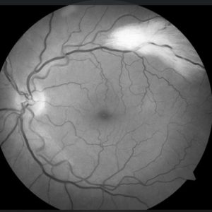
Photographic Imaging:
A color fundus photograph highlights the white myelination and feathery borders along the axons. Fundus autoflourescence (FAF) will show a dark appearance to the area of the myelinated RNFL. FAF works by detecting natural fluorescence, such as lipofuscin. The myelination appears dark in these situations where the myelination prohibits the detection of underlying fluorescent material (lipofuscin). A red-free image displays the white area of the myelinated RNFL. Myelin is mainly comprised of lipid, so red-free imaging is likely sensitive to areas high in lipid content. Lastly, OCT imaging demonstrates thickened RNFL in myelinated areas. It is assumed that the myelin creates bulk and thickness surrounding the axon; it is also assumed that the axon itself is larger as a result of the myelination. (2)
Differential Diagnosis: Myelinated nerve fiber layers are typically benign, but they can be mistaken for a variety of other retinal conditions. This retinal finding can be mistaken for a cotton wool spot, branch retinal artery occlusion, peripapillary epiretinal membrane, retinal pigment epithelium detachment, retinal infiltrate, retinoblastoma, or leukokoria. The most common differentials are cotton wool spots and branch retinal artery occlusion. (2)
References:
- Cheatham, K. M., Cheatham, M. A., Wood, K. B., & Kmk Educational Services, Llc. (2007). KMK Part One : Basic Science Review Guide (12th ed.). Overland Park, KS: Kmk Educational Services, Llc.
- Julie B. Shelton, MD. “Characteristics of Myelinated Retinal Nerve Fiber Layer in Opthalmic Imaging.” JAMA Network, 1 Jan. 2013, jamanetwork.com/journals/jama opthalmology/full article/1556904.
